Cervical Vertebrae (Cervical Spine)
Published on May 24th 2022 by staff
What is the Cervical Spine
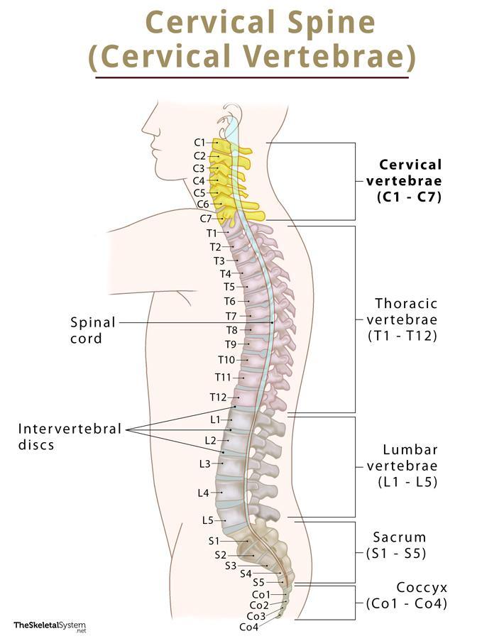
The cervical spine is the first part of the spinal column, consisting of 7 cervical vertebrae, C1-C7. These vertebrae are ring-like bony structures supporting the weight of the head. The first two bones, C1 and C2, are highly specialized, known as the atlas and axis.
Where are the Cervical Vertebrae Located
Cervical vertebrae are found in the neck region, starting from the base of the skull and extending to the thoracic cage of the trunk.
Quick Facts
| Type | Irregular bones |
| How many are there in the human body | 7 |
| Articulates with | Occipital bone, and 1st thoracic vertebrae |
Functions
- Support the head’s weight and allow a wide range of head and neck movements, like nodding and rotation.
- Protect the spinal cord as it passes through the hollow space encircled by the 7 bones.
- The C1 to C6 vertebrae contain small holes that Allow the vertebral artery, vein, and sympathetic nerves to pass through and carry blood to the brain. This is a unique feature of the cervical vertebrae as no other spine bones have such holes.
Anatomy and Structure of Bones in the Neck
Atlas and Axis
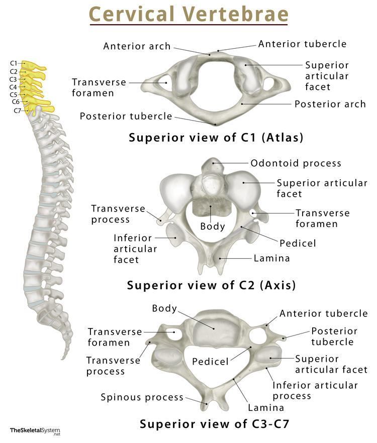
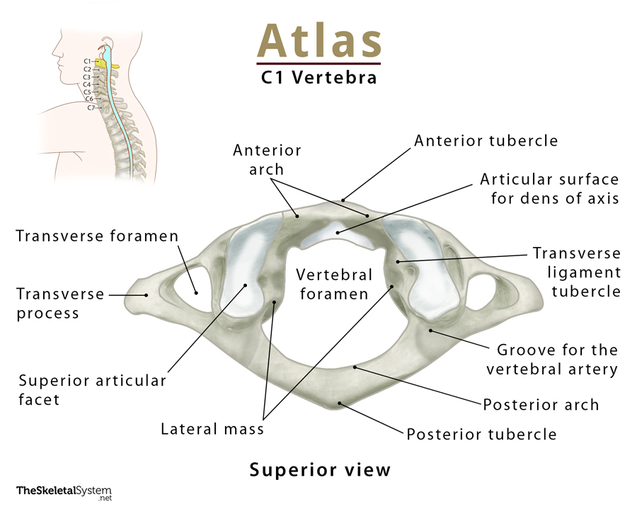
Atlas (C1): Located at the top of the spine, the atlas forms an articulation with the occipital bone, connecting the skull and spine. Its primary difference from the other vertebrae in the cervical region is the absence of a vertebral body and spinous process. Its lateral masses are connected by the anterior and posterior arches.
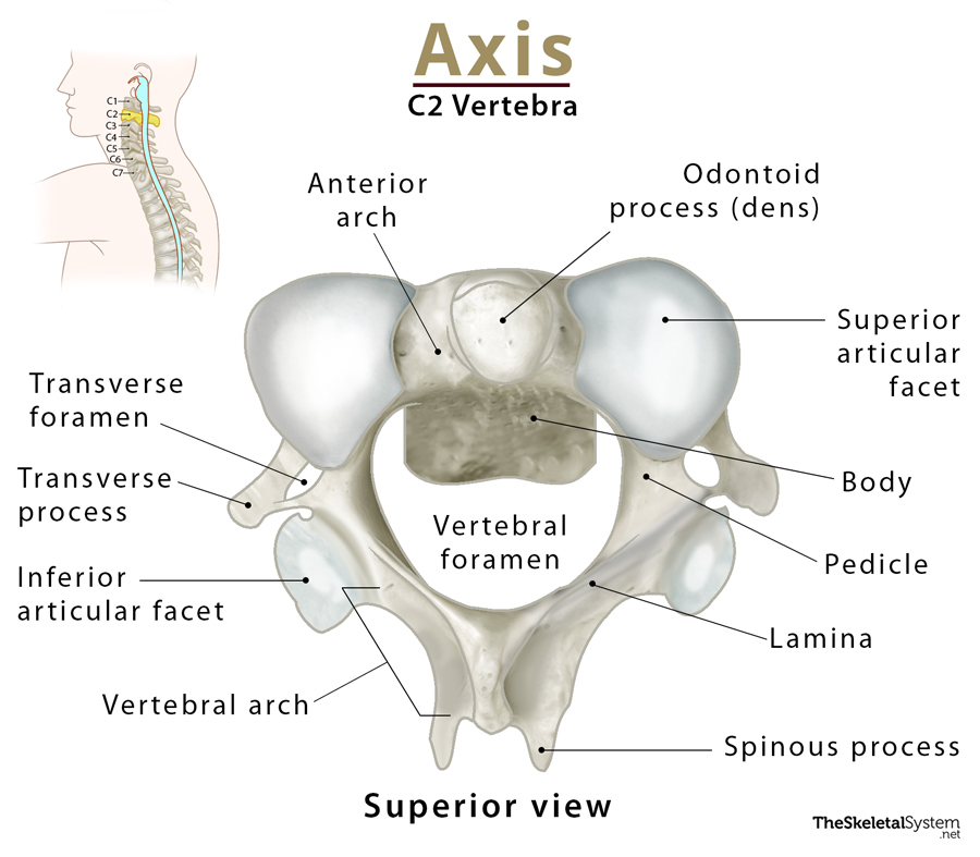
Axis (C2): It forms the pivot upon which the Atlas rotates (atlanto-axial joint), letting us rotate our head independently of the body. The most identifiable landmark of this bone is the strong odontoid process or dens that rises perpendicularly from the upper surface of the body and articulates with the atlas’ anterior arch.
There are also articulations between the axis’s superior articular facets and the atlas’ inferior articular facets.
Typical Cervical Vertebra (C3-C7)
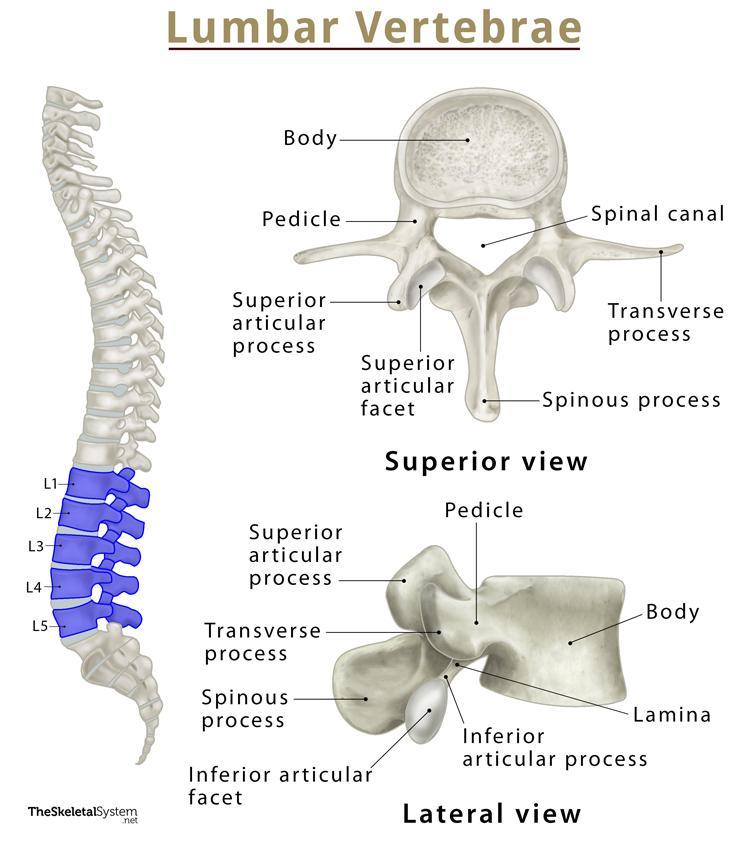
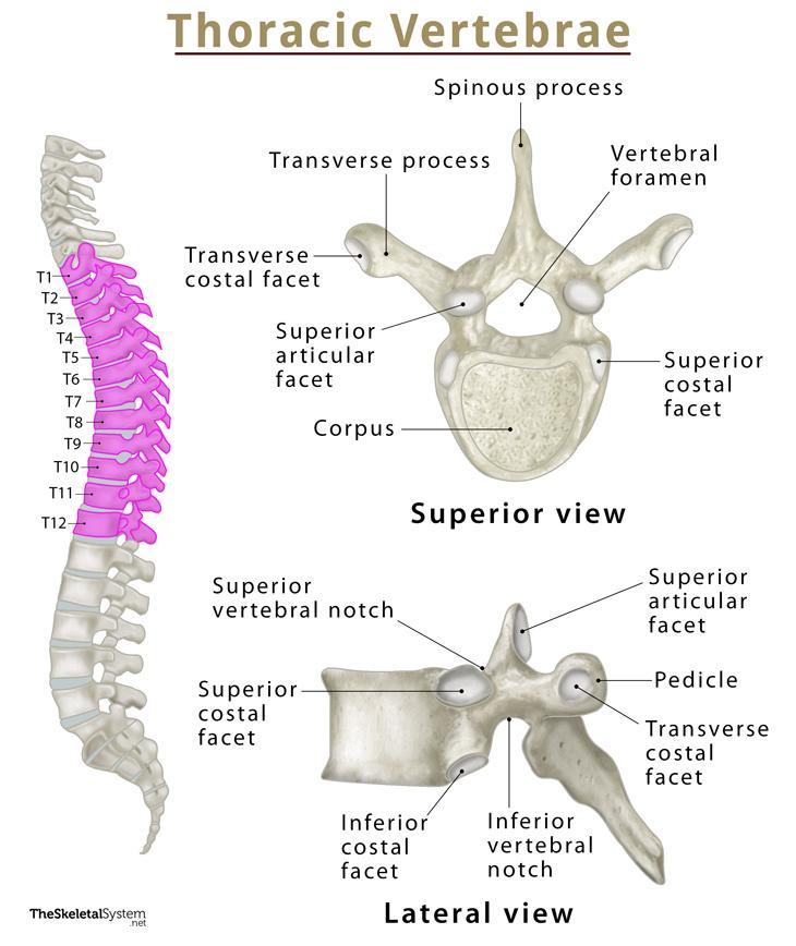
The next five vertebrae, C3-C7, have the typical structure for all the other vertebrae in the spine.
The thick, cylindrical part of each of these bones is called the vertebral body. This is the load-bearing part of the bone and is also the point for intervertebral articulation. An intervertebral disc lies at the place of articulation between two vertebral bodies. It provides cushioning and helps absorb any shock of movement at the joints.
The next part is the dorsal arch formed of two pedicles that arise at the dorsal part of the vertebral body. These two pedicles are joined by two flat laminae, which join at the midline to form the spinous process.
The dorsal side of the body, along with the pedicles and laminae, form a ring-like opening called the vertebral foramen. When the vertebra are stacked together, the vertebral foramen forms an opening through which the spinal cord runs. Protecting the spinal cord is the main purpose of this foramen. The place where the pedicles and laminae meet also has the superior and inferior articular processes, and transverse processes.
Articulations
The vertebrae articulate with each other through facet joints, and there are the three following joints in the cervical spine region:
- Atlanto-occipital joint: The synovial joint formed between the atlas (C1) and occipital bone.
- Atlanto-axial joint: The joint formed by the articulation between the atlas and axis.
- Uncovertebral joints: Small synovial joints between the five typical vertebrae.
Muscle and Ligament Attachments
Muscle attachments:
- Lumbar muscles
- Thoracic muscles
- Back muscles
Ligament attachments:
References
- The Cervical Spine — Teachmeanatomy.info
- Anatomy, Head and Neck, Cervical Vertebrae — Ncbi.nlm.nih.gov
- Cervical spine — Kenhub.com
- Cervical Vertebrae — Innerbody.com
- Cervical Vertebra — Sciencedirect.com

